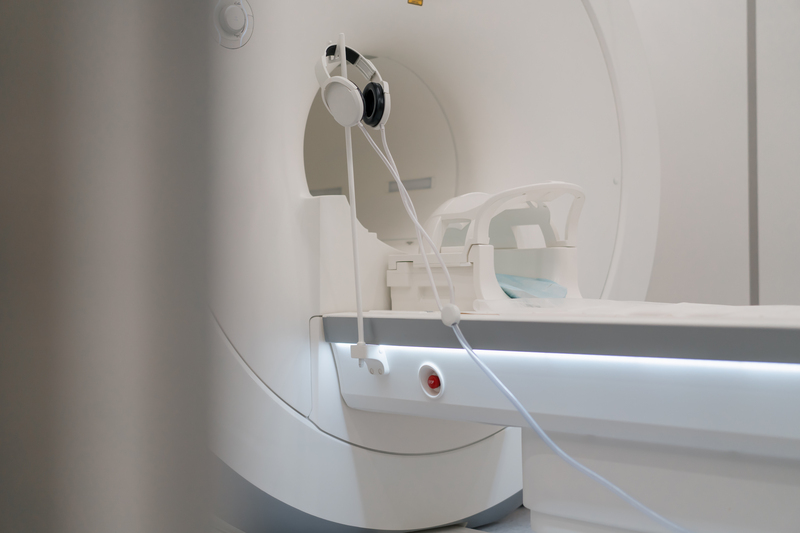
UroNav was introduced in 2013 by Invivo, a division of Philips Medical Systems. They devised a mechanism that enables an MRI to be superimposed over the transrectal ultrasound image. When this “Fusion” of images occurs, the abnormal areas in the prostate can be visualized.
When the Multi-Parametric MRI image is fused to the ultrasound image, the urologist is able to visualize the MRI-identified abnormality and direct the needle with ultrasound guidance. This is important for several reasons:
- The standard template or map that is used with non-fusion guided biopsies does not sample the anterior or front part of the prostate
- First time non-fusion ultrasound guided biopsies miss 25-30% of prostate cancers
- Many of the more aggressive prostate cancers are found in the anterior part of the prostate
- Abnormal areas in the remainder of the prostate can also be visualized and targeted for biopsy
- Utilizing MRI-Fusion technology, abnormal areas in the anterior part of the prostate can be visualized and reliably targeted and biopsied
Should everyone who needs a prostate biopsy have an MRI-Fusion biopsy? NO! For patients with an elevated PSA or abnormal area in the prostate that can be detected by digital exam, and who are thought to be at risk for having prostate cancer, non-fusion transrectal ultrasound guided prostate biopsy is certainly reasonable.
MRI-Fusion biopsy should be considered by the following patients:
- Patients who have had a negative ultrasound-guided prostate biopsy and in whom prostate cancer is still suspected
- Persistent, unexplained elevated PSA
- Increased prostate cancer gene expression such as PCA3
- Patients who are part of an Active Surveillance protocol (MRI-Fusion biopsy is now standard in the largest Active Surveillance program in the US and the world)
For more information, contact our office at Vitality Plus Urology today!
